Personal Blogs This mini size heart is also
Illustrated anatomy of the heart: illustrations and photography. Some illustrations have been drawn and coloured with Adobe Photoshop. Heart - Human anatomy : Cross sections (Right/left ventricle, Right/left atrium, Interventricular septum Others are based on an adapted 3D model (texturing and modelling) to the study of anatomy.

Anatomical Heart Sketch at Explore collection of
Hello scientist! This video shows you how to draw a heart for your scientific figure and graphical abstract. 🎨 DrawBioMed is a channel for scientists to lea.

Pin on Wedding Look
With this easy human heart drawing ideas, you can learn how to draw a human heart easily. I made this cool drawing as a guide for you to create a simple anat.

Corazón. Heart anatomy, Human anatomy art, Medical illustration
The heart has three layers. They are the: Epicardium: This thin membrane is the outer-most layer of the heart. Myocardium: This thick layer is the muscle that contracts to pump and propel blood.
Anatomy Of The Heart Clipart Human Heart Drawing Png, Transparent Png
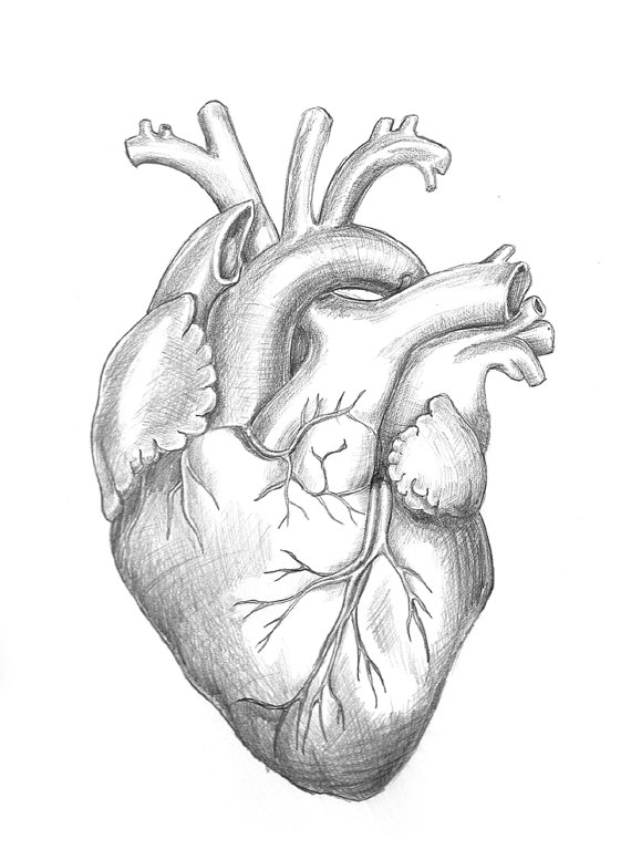
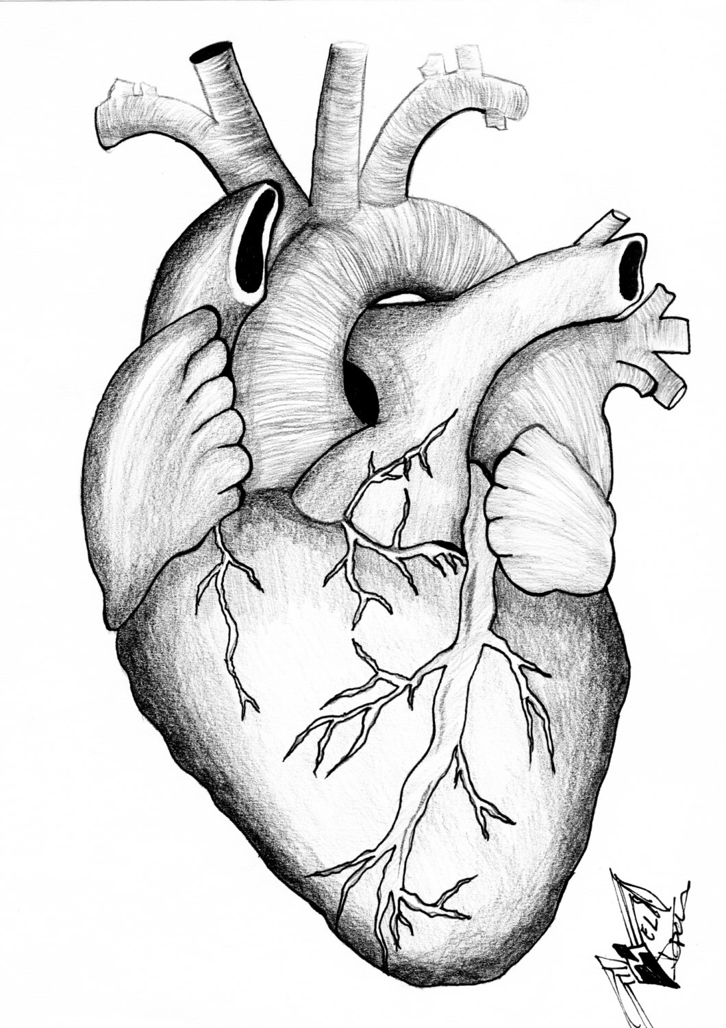
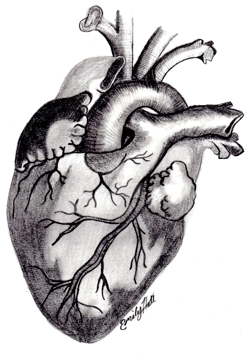
Color Human Heart Drawing . Above is a medical diagram of an Anatomical Heart Drawing from an old Nurses Anatomy book. It is a full color Heart Illustration with blue and red sections along with black and white. It is an anatomically correct heart drawing illustrating valves, veins, vessels, ventricles, chambers, etc.

Den inneren Aufbau des Herzens zeichnen (mit Bildern) wikiHow
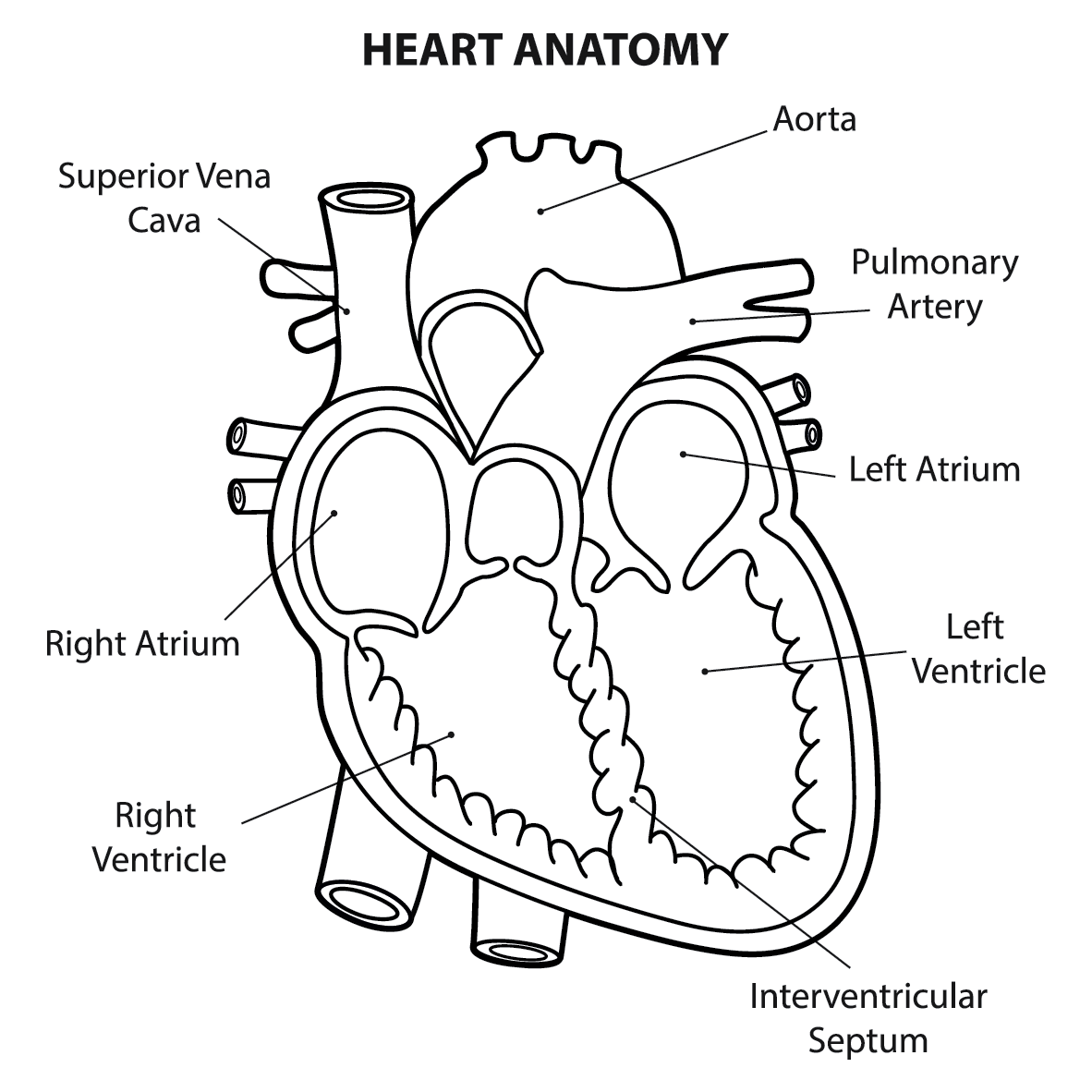
Location of the Heart. The human heart is located within the thoracic cavity, medially between the lungs in the space known as the mediastinum. Figure 19.2 shows the position of the heart within the thoracic cavity. Within the mediastinum, the heart is separated from the other mediastinal structures by a tough membrane known as the pericardium.
Anatomical Drawing Heart at GetDrawings Free download
1. Draw a tilted and irregular curved shape in the center of your page. Use a pen or pencil to draw the heart's main body. Create a curved shape similar to an acorn or apple's bottom half. Angle the slightly tampered end of the shape to the left about 120 degrees. [1]
Anatomical Heart Pictures Cliparts.co
Function and anatomy of the heart made easy using labeled diagrams of cardiac structures and blood flow through the atria, ventricles, valves, aorta, pulmonary arteries veins, superior inferior vena cava, and chambers. Includes an exercise, review worksheet, quiz, and model drawing of an anterior view (frontal section) of the heart in order to.
anatomicaldrawingheart5 • Heritage Direct Primary Care
Drawing the heart anatomy can be challenging, but with practice and dedication, it can become easier over time! The human heart is a complex organ with many parts and intricate details, which can make it difficult to draw accurately. It requires a good understanding of proportions and shading to create a realistic depiction.

How to Draw a Human Heart 11 Steps (with Pictures) wikiHow
Heart structure & function, conduction, congestive heart failure, erythropoiesis. Videos, follow-along-notes, practice questions.. Quizlet Basic Heart Anatomy. Sympathetic Stimulation of the Heart. Drawing to follow along with me; Typed Lecture Notes; Practice Questions (and Answers)
Anatomical Drawing Of The Heart at GetDrawings Free download
To draw an anatomical heart requires a lot of attention and patience. Perfect the pencil drawing before moving on to the pen. This will make the pen drawing process much easier as we use the pencil marks for guidance. Take a break. The tutorial can be done in sections, so perhaps take a break if you need to.
Vintage Anatomical Heart Drawing Free download on ClipArtMag
Now that we have a basic understanding of the heart's anatomy, let's get started on drawing it! Begin by drawing a large oval shape for the main body of the heart. Next, draw a smaller oval shape at the top of the heart for the left atrium. Draw a curved line connecting the left atrium to the main body of the heart.

thelexirose Just another site
Heart anatomy. The heart has five surfaces: base (posterior), diaphragmatic (inferior), sternocostal (anterior), and left and right pulmonary surfaces. It also has several margins: right, left, superior, and inferior: The right margin is the small section of the right atrium that extends between the superior and inferior vena cava .
Human Heart Drawing Images at GetDrawings Free download
In this lecture, Dr Mike shows the two best ways to draw and label the heart!
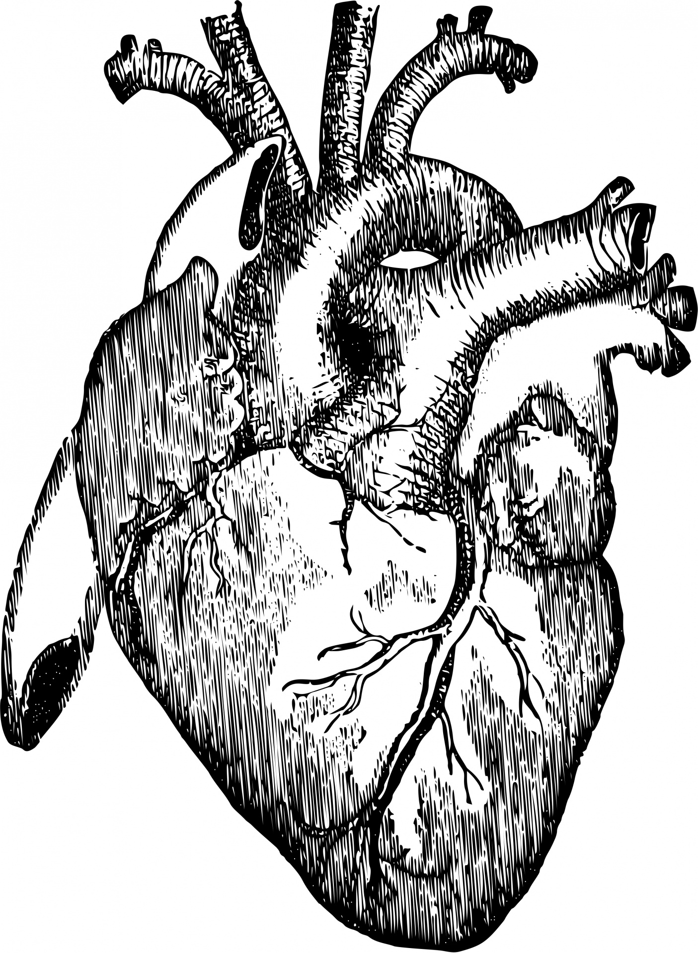
Vintage Anatomical Heart Drawing at GetDrawings Free download
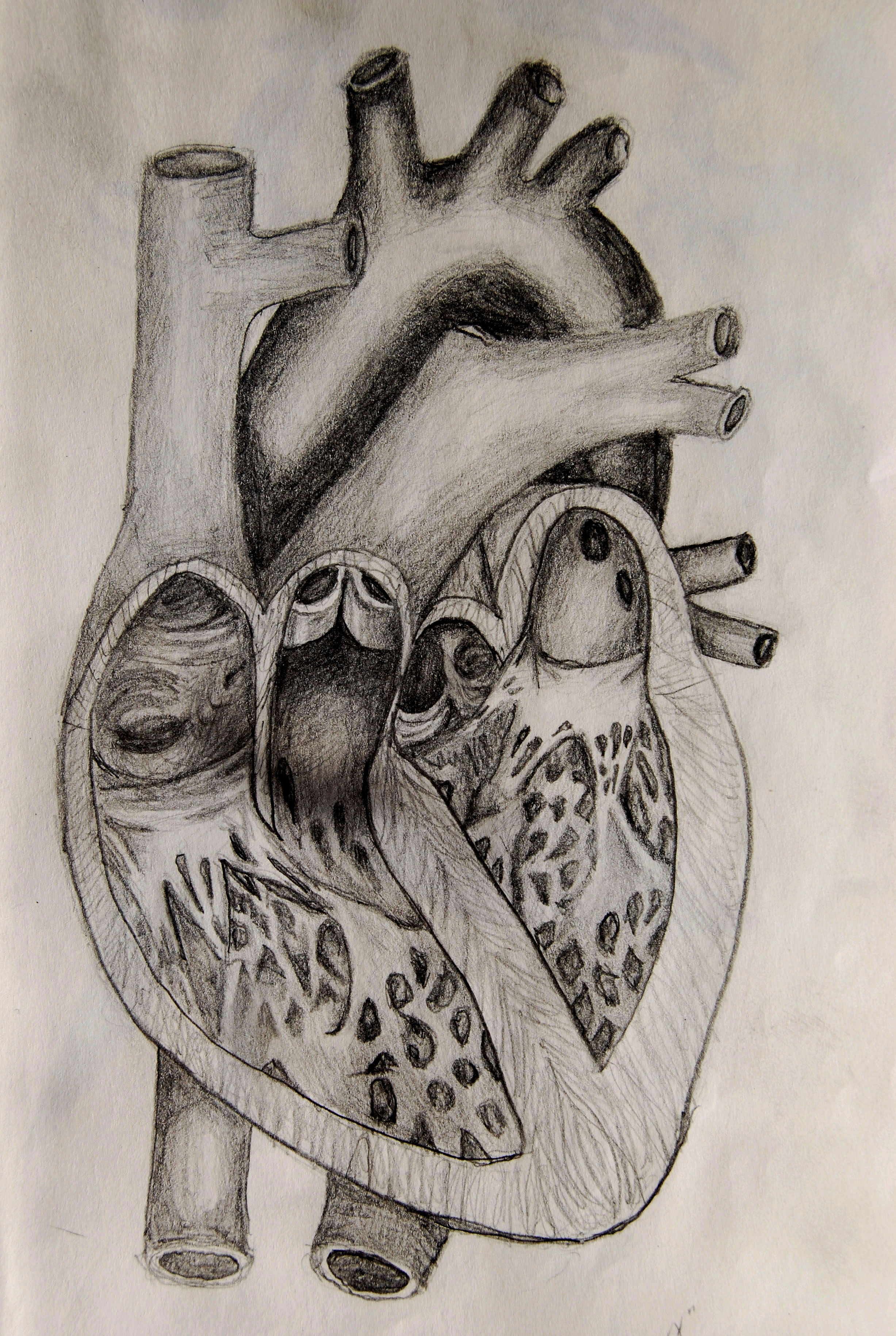
Drawing Internal Anatomy of the Heart. A drawing of the anatomy of the opened normal heart, with English labels. From the OpenStax Anatomy and Physiology book. Valves. Video Beating Aortic Valve. A video of the aortic valve in a cadaver heart that is beating in a laboratory set-up by the University of Minnesota.
Human Heart Anatomy Drawing at GetDrawings Free download
Step 2: Add Details. Add the pulmonary arteries to the triangle shape, one on each side. Then, draw the aorta coming out of the top of the heart and curving upwards towards the neck. Draw the superior and inferior vena cava coming into the heart from the top and bottom. Don't forget to add a small right auricle too!
